 |
| |
Viet Nam News
by Dr. Christian Brosset*
What is an electromyogram?
Electromyogram (EMG) is a diagnostic procedure to evaluate the health and functioning of muscles and nerve cells that control them. These nerve cells are called moto neurons and they send electrical signals to the muscle, which reacts by contracting. An EMG translates these signals into graphs and numerical values that your neurologist can read and interpret. Neurologists consider the EMG an extension of the physical examination of the patient and it is the gold standard for the diagnosis of numerous disorders affecting the peripheral nervous system.
How is it done?
The examination is divided into two parts and takes approximately 45 minutes. No special preparation is required. The first part is a nerve conduction study (NCS), which uses electrodes taped to the skin to measure the speed and strength of the electrical signal travelling along your nerves and muscles. The second part is a needle EMG. A tiny needle is inserted into your muscle and the electrical activity in the muscle is recorded. The needle EMG may not be required for everyone; it can be performed in the outpatient department and you can go home afterwards.
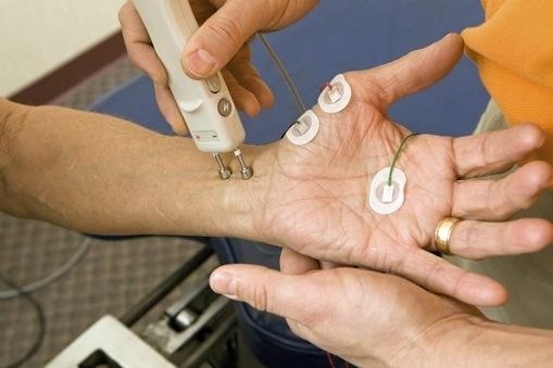
- Nerve Conduction Study (NCS)-Stimulation Detection: A recording electrode will be placed on your skin, usually on your arms and/or legs to obtain measurements of your nerve impulses and the way in which muscles react to them. Another electrode will be used to stimulate the nerve. The stimulator produces small electrical pulses, which feel like a sharp tapping sensation. The process may be repeated for a number of nerves. A NCS measures the speed for a nerve impulse to travel along a nerve and how muscles respond to the signal. If the nerve is trapped, damaged or diseased then these signals will be slow and/or the amplitude of your muscular response, flatter.
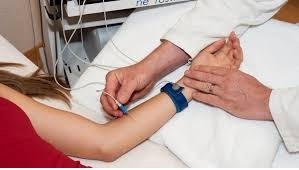
- Needle EMG-Detection: During a needle EMG, the electrical activity of your muscles is measured at rest and after muscle contraction, which is directly recorded by inserting a small needle electrode into your muscle(s). The needles are different and smaller than the needles used for injection of medications, so discomfort is much less. This part of the examination is necessary for the diagnosis of conditions such as sciatica or cervico-brachial neuralgia. For such conditions, the NCS will show normal readings and they can only be detected by needle EMG. It is also the gold standard for the detection of all muscle pathologies, such as myopathies, myositis, or muscular dystrophies.
Why is it done?
The EMG is used in the exploration of the “peripheral nervous system” (PNS). It refers to the part of the nervous system outside the brain and the spinal cord (called the central nervous system or CNS) and consists of the nerves, plexus and roots. Its function is to connect the brain and spinal cord to the muscles and the rest of the body. Your doctor may order an NCS and/or EMG if you have signs or symptoms that may indicate a nerve or muscle disorder including:
- Tingling
- Numbness
- Muscle weakness
- Muscle pain or cramping
- Certain types of limb palsy
Although some conditions only require an NCS to reach a diagnosis, needle EMG results are often necessary to help diagnose or rule out a number of conditions such as:
- Disorders of nerves outside the spinal cord (peripheral nerves), such as carpal tunnel syndrome (CTS) or peripheral neuropathies. CTS is very common among manual workers and pregnant women and can be associated with general conditions such as hypothyroidism. It is caused by the compression of the median nerve during its passage under the carpal ligament through your wrist (carpal tunnel). CTS may cause pain, numbness and a tingling sensation in the thumb, index and middle finger as well as the thumb side of the ring finger. The EMG will provide the diagnosis and the decision whether the CTS needs to be treated by surgery or not.
- Disorders that affect the motor neurons in the brain or spinal cord, such as amyotrophic lateral sclerosis or polio.
- Traumatic nerve injury. After an accident or injury, the EMG can detect the location and extent of a nerve damage and provide a prognosis of the recovery time.
- Disorders that affect the nerve root, such as a herniated disk in the spine, which can cause painful nerve root compression such as Sciatica. It is characterised by a very sharp pain originating in the lateral region of the lower back radiating to the lower limb and back of the foot or heel. The EMG can help determine whether surgery or conservative treatment is necessary.
- Muscle disorders, such as muscular dystrophy or polymyositis. The EMG together with a muscle biopsy will be able to detect such conditions.
- Diseases affecting the connection between the nerve and the muscle, such as myasthenia gravis.
- Peripheral neuropathy, which refers to the damage or impairment of the PNS as part of a generalised condition. Diabetes, chronic alcohol poisoning and certain drugs such as chemotherapeutic agents for the treatment of cancers can damage your peripheral nerves and cause polyneuropathy. Symptoms may include weakness, numbness and pain, usually in your hands and feet. It can also affect other areas of your body.
* Dr. Christian Brosset works at the Neurology Department at HFH and has brought with him his long-term expertise in disease prevention and treatment. He is a specialist in EMG and treatment of PNS disorders.
If you have any questions or want to book an appointment with our doctors, please contact us on our phone number 84 – 24.3577.1100, access www.hfh.com.vn, or email us at contact@hfh.com.vn, Address: 1 Phuong Mai, Dong Da, Ha Noi
 |
| Needle EMG-Detection. — Photo courtesy of Hanoi French Hospital |
 |
| Nerve Conduction Study (NCS)-Stimulation Detection. — Photo courtesy of Hanoi French Hospital |
 |
| Dr. Christian Brosset. — Photo courtesy of Hanoi French Hospital |
OVietnam